The Pupillary Light Reflex is very important in the neurological assessment, as it provides first-rate information regarding the CNS. This includes the reflex in which the pupil of the eye constricts or dilates in response to light and is underpinned by complex neural networks. Abnormalities in this reflex indicate neurological problems. Neurological Pupil Index (Npi) is useful in measuring these oddities. Here’s an overview of the typical patterns of pathological changes in the pupil’s reaction and the significance of these changes in clinical practice.
A Review of the Pupillary Light Reflex
Pupillary light reflex refers to the following neural circuits. When light reaches the eye it is perceived by the retina and an electrical impulse is sent to the optic nerve which in turn transmits the message to the pretectal nucleus in the midbrain. It goes to the Edinger- Westphal nucleus and then to the oculomotor nerve that supplies the iris sphincter muscle which leads to constriction of the pupil. This reflex is essentially performed during a neurological examination to evaluate the status of these pathways.
Neurological Pupil Index (Npi)
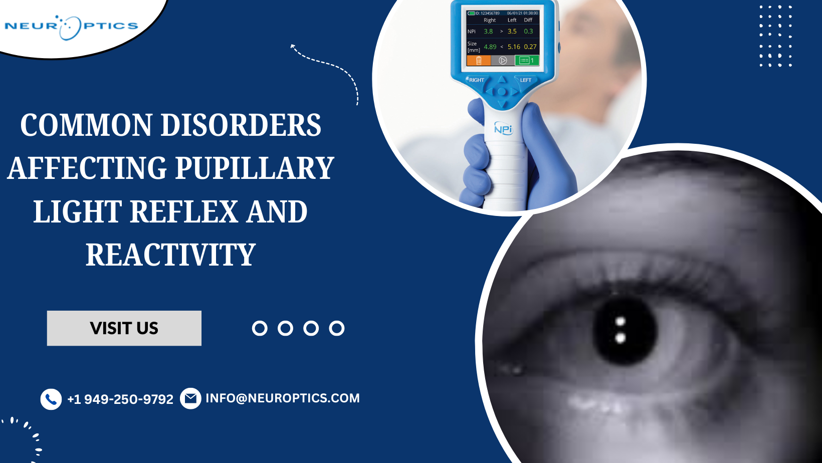
The Neurological Pupil Index (Npi) is a quantitative measure used to evaluate pupil reactivity. This standardized score is based on pupil size, constriction velocity, and dilation velocity, offering a precise gauge of neurological function. Npi values range from 0 to 5, with lower scores indicating impaired pupillary response.
Common Diseases That Influence Pupillary Light Reflex
Optic Neuropathies
Optic neuropathies, such as optic neuritis or ischemic optic neuropathy, disrupt the afferent pathway of the pupillary light reflex. Patients with these conditions often present with a relative afferent pupillary defect (RAPD), where the affected eye shows reduced constriction compared to the unaffected eye when exposed to light.
Optic Neuritis: Optic Neuritis is often linked with multiple sclerosis Optic neuritis is a condition that results in inflammation of the optic nerve which causes visual disturbances and also affects the pupillary reflex.
Ischemic Optic Neuropathy: It is a condition common with elderly individuals caused by occlusions of the optic nerves. It presents with asymptomatic, monocular, and sudden loss of vision and RAPD.
Oculomotor Nerve Palsy
Injury to cranial nerve III affects the afferent pathway of the Pupillary Light Reflex and results in a dilated pupil that doesn’t constrict in response to light.
Aneurysms and Tumors: Oculomotor nerve palsy due to cerebral aneurysms or tumors can be common and a blown pupil (severe mydriasis) is a key diagnostic sign.
Diabetic Neuropathy: Diabetic microangiopathy is a common cause that can affect the oculomotor nerve causing similar complaints.
Horner’s Syndrome
Horner’s syndrome is a clinical syndrome involving ptosis (drooping eyelids) accompanied by miosis (pinpoint pupil) and anhidrosis (absence of sweating), which occurs due to damage to the sympathetic pathways to the eye.
Central Lesions: Horner’s syndrome can be caused by hypothalamic or brainstem strokes or tumors along with the cervical spinal cord.
Peripheral Lesions: Other causes include trauma to the sympathetic chain or tumors in the neck that compress the carotid artery, or carotid artery dissection.
Adie’s Tonic Pupil
Adie’s tonic pupil is an asymmetrical one in which one of the pupils is larger than the other and the slow pupillary response to light but brisk near reflexes.
Idiopathic Etiology: The cause of Adie’s tonic pupil is unknown in many cases but it is suggested that the tonic pupil arises from damage to the postganglionic fibers of the parasympathetic supply.
Holmes-Adie Syndrome: However when accompanied by absent deep tendon reflexes it is known as Holmes–Adie syndrome.
Pharmacologic Influence
There are known drugs and chemicals that can alter how the pupillary light reflex as well as reactivity.
Mydriatics and Miotics: Atropine can lead to a state of pupillary dilatation and pilocarpine can cause pupillary constriction.
Opioids: Opioid-induced intoxication presents with miosis.
Diagnostic Tools and Techniques
Neurological Exam
To this date, a comprehensive neuro exam continues to be the best technique for evaluating the PLR. This is achieved by observing the pupils in size, shape, and contraction to light using a flashlight or pen light.
Automated Pupillometry
Computerized pupillometry equipment facilitates the quantification of the size of the pupils and their reaction, which yields the Npi. It improves the reliability and validity of the pupillary measurements, especially for patients in the intensive care unit.
Neuroimaging
MRI and CT scans are crucial in assessing the structural damages influencing the nuclei and fibers responsible for the PLR. These modalities are more applicable in the diagnosis of diseases such as tumors, aneurysms, and stroke.
Clinical Management and Implications
Effective management of disorders affecting the pupillary light reflex is essential for optimal patient outcomes. Early diagnosis and intervention are critical.
Optic Neuropathies
Management: Treat optic neuritis with corticosteroids. Manage ischemic optic neuropathy by controlling vascular risk factors like hypertension and diabetes.
Prognosis: Optic neuritis generally has a good prognosis. Ischemic optic neuropathy can lead to permanent vision loss.
Oculomotor Nerve Palsy
Management: Immediate neuroimaging is crucial to rule out aneurysms. For diabetic neuropathy, maintain strict glycemic control. Surgical intervention may be needed for compressive lesions.
Prognosis: Diabetic palsies often improve on their own, while aneurysmal compression may require surgery.
Horner’s Syndrome
Management: Treat the underlying cause, which may involve surgery for tumors or management of carotid artery dissections and strokes.
Prognosis: Depends on the cause, with possible partial or complete resolution.
Adie’s Tonic Pupil
Management: Symptomatic treatment includes reading glasses and pilocarpine eye drops.
Prognosis: Generally benign with a favorable outcome.
Pharmacologic Influence
Management: Discontinue the offending drug. Use naloxone for opioid intoxication. Adjust other drug dosages as needed.
Prognosis: Good, with pupils returning to normal once the drug is stopped.
Research and Future Directions
The research process of the PLR and its disorders is still progressing. Such studies seek to enhance the current and future diagnosis, treatment, and general well-being of patients.
Innovative Diagnostic Tools
Better technologies in the pupillometry equipment and computer programs used for the measurements will improve the accuracy of pupil reactivity. Such changes are believed to offer better and prior identification of neurological conditions.
Molecular and Genetic Insights
Adie’s tonic pupil and Horner’s syndrome may be well explained at the molecular and genetic levels; thus, subsequent research focusing on these levels could reveal new therapeutic avenues. There could be genetic markers that doctors could identify and use to devise custom-made treatments.
Neuroprotective Therapies
New therapy approaches that shield and restore the neural circuitries related to the Pupillary Light Reflex have potential. Neuroprotective agents or pharmacological therapies and regenerative medicine like stem cell therapy are in active exploration.
Conclusion
This test is also a vital part of the neurological examination since allows us to evaluate the condition of the central nervous system. This reflex can be altered by various disorders including optic neuropathies, oculomotor nerve palsy, Horner’s syndrome, Adie’s tonic pupil, as well as pharmacologic interference and each case needs to be handled appropriately. Automated pupillometry and neuroimaging are also essential in the assessment and diagnosis of these disorders, with the Neurological Pupil Index (Npi) being one of the most significant neurological tools in this process.
The first signs and proper management are key factors in the best way to help the patient. Future research and development will that add to the knowledge and improve ways of diagnosing and treating the disorders. Clinicians should not overlook the tests that involve the evaluation of the pupillary light reflex and its reactivity, as these tests give very important information about the overall neurological status of a patient.